International Journal of Veterinary Science and Research
Surgical correction of omphalocele in local goat breed (Beetal) of Jhang, Punjab: A case study
Ali Raza1*, Wajeeha Saeed1, Abdul Mateen1, Aun Muhammad1, Farah Ijaz1 and Amanullah Khan2
2Department of Pathobiology, College of Veterinary and Animal Sciences, Punjab, Pakistan
Cite this as
Raza A, Saeed W, Mateen A, Muhammad A, Ijaz F, et al. (2023) Surgical correction of omphalocele in local goat breed (Beetal) of Jhang, Punjab: A case study. Int J Vet Sci Res 9(3): 059-62. DOI: 10.17352/ijvsr.000138Copyright License
© 2023 Raza A, et al. This is an open-access article distributed under the terms of the Creative Commons Attribution License, which permits unrestricted use, distribution, and reproduction in any medium, provided the original author and source are credited.Omphalocele is a rare congenital condition where closure defects in the abdominal wall at the umbilical ring lead to the protrusion of intestinal or other visceral organs, covered by a thin epithelial layer. The developmental mechanism of this condition is not fully understood, and various theories have been proposed to explain it. This study presents a case of omphalocele in a newborn female black goat kid, detailing its clinical presentation, surgical management, and postoperative care. The surgical procedure involved meticulous preparation of the surgical site, administration of local anesthesia, and careful repositioning of the intestines, liver, and a portion of the spleen. Excess skin and the amnion membrane were removed to facilitate safe repositioning, and the umbilical ring was excised to widen the opening. The abdominal wall layers were meticulously closed using appropriate suture materials. The kid’s postoperative recovery was uneventful, with normal vital signs, fecal passage, and feeding behavior observed. The study discusses omphalocele in comparison to other abdominal abnormalities and explores potential developmental mechanisms. The authors emphasize the importance of immediate surgical intervention despite varying prognoses associated with this condition. The study underscores the significance of surgical treatment for omphalocele cases in newborn goat kids, providing hope for affected animals and valuable insights for veterinary professionals. Although the exact prevalence of omphalocele remains uncertain due to unreported cases, this report demonstrates successful surgical correction and the potential for curing the condition if diagnosed and treated promptly. Further research is needed to fully understand the genetic and environmental factors contributing to omphalocele and its impact on livestock.
Introduction
Omphalocele is a rare congenital condition with closure defects in the abdominal wall at the site of the umbilical ring. This defect causes the protrusion of the intestinal or other visceral organs, which are covered by a thin layer of epithelium [1]. The exact process of development of this defeat is not fully understood. However, different theories have tried to explain its mechanism of development. Embryonic dysplasia can occur due to any disruption in ectodermal placodes (normally present at the umbilical ring) during embryonic life [2]. This abnormality was regarded as the cause of failure of body wall folding and closing of the abdominal wall, causing enlargement of the umbilical ring [3]. Another theory states that this abnormal condition occurs during embryonic development when there is a failure of normal migration of one of the four body folds [4]. The inability of the herniated part during embryonic life can also lead to the development of the omphalocele [5]. The genetic link to this condition is not fully understood. However, some authors have considered it as an inherited condition, which may be linked to factors like inbreeding [6]. In humans, the abnormal karyotype is present in almost 40% of patients with omphalocele [7]. It is difficult to determine the true prevalence of this condition due to unreported deaths [8]. The size of the opening and the areas involved determine its prognosis. However, it has a good prognosis if treated well on time and with precision [9]. Different environmental factors are also associated with this congenital defect. Many other factors like nutrition, drugs, and different toxins can also play their part in these disorders [10]. A short gestation period can act as a risk factor. The ratio of these congenital disorders ranges from 2-3:1000 in cattle, sheep, goats, and buffalo. Emergency surgery is indicated for a case of omphalocele to save the life of the animal. This condition is not breed-specific.
Case history
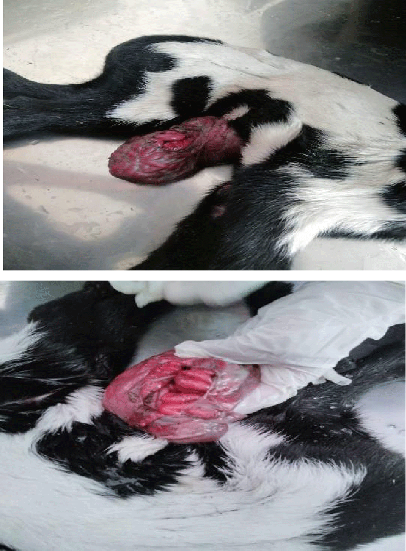
A newborn female, black goat kid, local breed (Beetal), weighing 3 kg, was presented at the veterinary teaching hospital, College of Veterinary and Animal Sciences, Jhang, with evisceration of the intestines through the umbilical ring from the ventral abdomen (Figures 1,2). The kid was normal, full-term, and the birth was unassisted. Rectal temperature, heart rate, and respiration rate were normal. The intestines in the form of soft pendulous sacs, were hanging through the umbilical ring from the ventral abdomen, which measured between 1.75-2.5cm. A clear thin membrane (amnion) around this sac was ruptured, slightly congested, and contaminated with manure and dirt. Some intestinal loops in the sac were normal. The pendulous mass was covered in a clean cloth by the owner. The umbilical cord was not connected to the ring. The umbilical ring had well-developed boundaries. The history and clinical assessment suggested that it was a case of omphalocele. A reconstructive surgery (Herniorrhaphy) was recommended as a treatment.
Surgical management
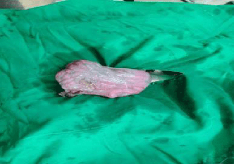
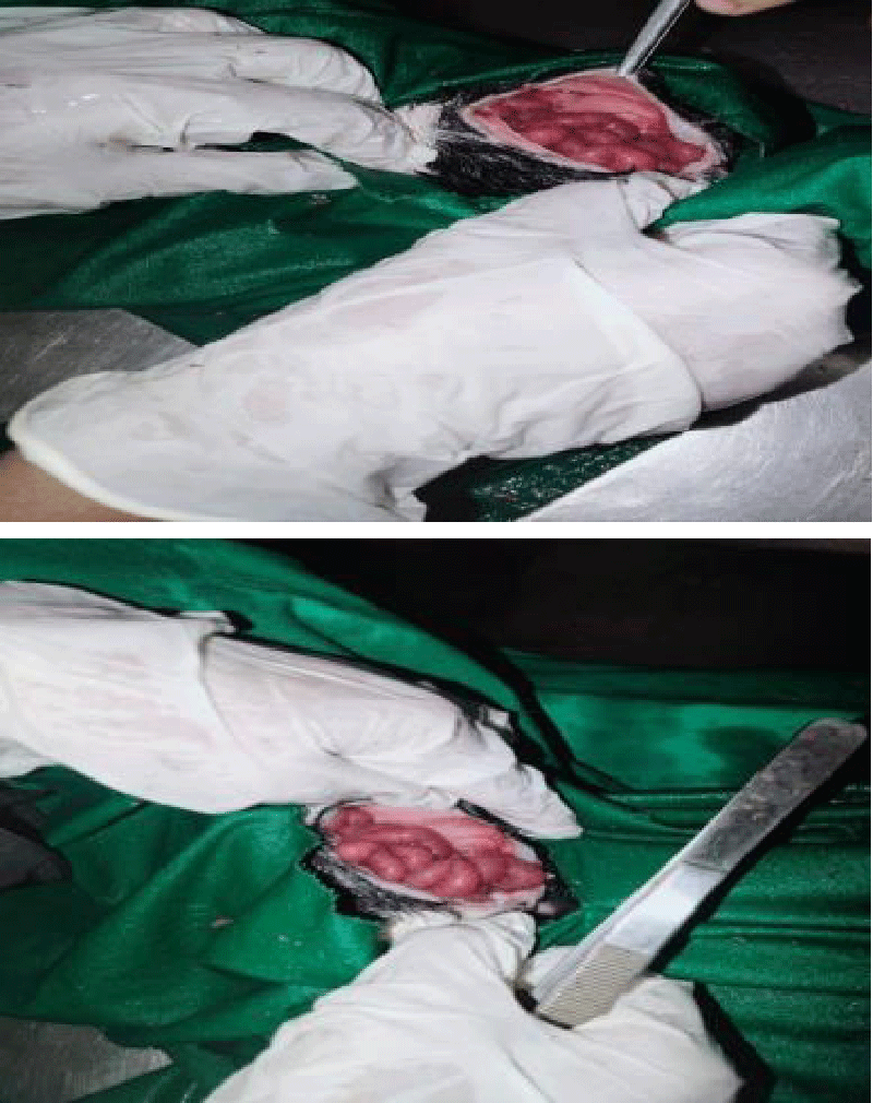
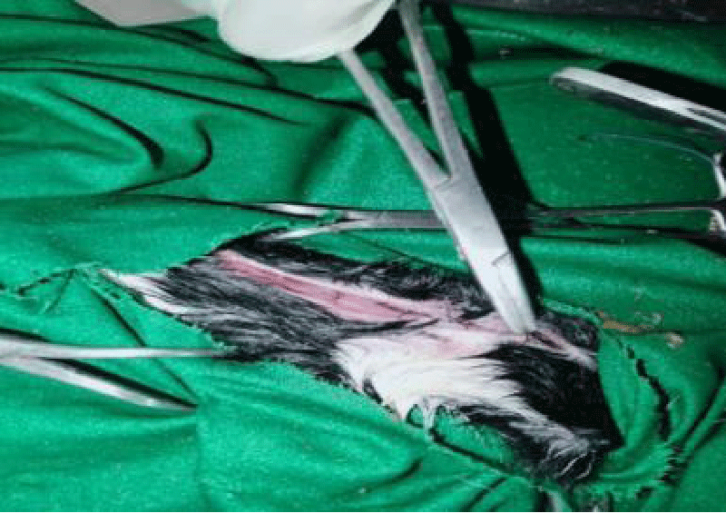
To perform the surgical procedure for correction of omphalocele, the surgical site around the umbilical ring was meticulously prepared using sterile techniques (Figure 3). Lidocaine HCL (Xyloid® 2%) was administered as local anesthesia via a ring block to ensure pain relief during the procedure. Lukewarm normal saline was used to flush and wash the protruding intestines, contaminated with manure and dust. Due to the risk of potential bleeding, some dirt attached to the intestines was left untouched. To facilitate the repositioning of the bowel, excess skin, and the amnion membrane were carefully removed. The umbilical ring was excised to widen the opening, allowing for the safe relocation of the intestines. The skin incision was extended (approximately 3 cm) cranially, and liquid paraffin was applied to lubricate the intestines, ensuring a gentle and injury-free repositioning process (Figure 4,5).
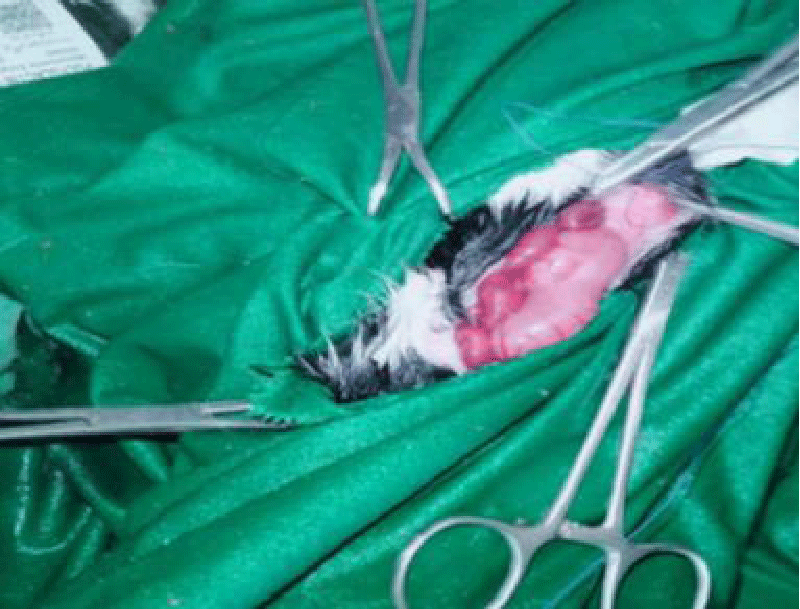
Various suture materials were used to close the layers of the abdominal wall. An absorbable suture material 2 - 0 (Chromic catgut®) was utilized for suturing the peritoneum and a thin layer of muscle using cross mattress suture pattern (Figure 6). The skin was sutured using a simple interrupted pattern with silk braided No. 1-0 (Silk®) (Figure 7). Throughout the surgical procedure, the vital signs of the goat kid, including temperature, pulse rate, and respiratory rate, were closely monitored and found to be within the normal range. The eviscerated part of the intestine was evaluated for its healthy color and peristaltic movement after being gently placed back into the abdominal cavity.
Following the surgery, no complications were noticed. The owner was guided to offer the dam’s milk to the kid for proper nutrition. The kid experienced normal fecal passage, and there were no gastrointestinal issues throughout the recovery period.
Postoperatively, a combination of skin ointment (Polyfax®) and Procaine Penicillin (50,000 IU/kg/day) was prescribed with a proper dressing of the wound. The kid was examined after a week and was found normal. Suture line and feeding behavior were normal. After two weeks, the wound was almost healed and sutures were removed. The kid had a normal fecal passage with no GIT problems.
Discussion
A common region for the occurrence of congenital abdominal abnormalities is the umbilicus. Congenital umbilical hernia can occur through incomplete closure of the abdominal wall at the umbilical area [11]. Similarly, gastroschisis and omphalocele also occur through defects in the umbilicus [12]. Protruded area and herniated parts from the umbilicus were the main points for the diagnosis of this case. An omphalocele has a covering of a membrane composed of two layers, an outer amnion, and peritoneum. In some cases, Wharton’s jelly, a gelatinous connective tissue was found in the umbilical region [13]. In some cases, in lambs, loops of intestines were found within the herniated portion [1]. In cattle calves, the sac can have intestines with a small portion of the liver. However, in goats, intestines are presented along with the liver and a portion of the spleen [14].
In omphalocele, there is an evisceration of a part of the small intestine, large intestine, and other portions of the gastrointestinal tract, like the liver [15]. Gastroschisis is the major condition that should be differentiated from omphalocele. In Gastroschisis, the umbilical cord is not involved and the protruded mass is usually to the right of the midline. Another distinguishing point of the two conditions is the presence of a membranous sac, which is present in the case of gastroschisis [16]. Other two different abnormalities are schistosomus reflexus and congenital umbilical hernia. In congenital hernia, eviscerated organs are covered with skin. In schistosomus reflexus, there is retroflexion of the vertebral column, and ankylosis of the axial skeleton and limbs in addition to the evisceration of intestinal organs (Dennis and Meyer, 1965).
A normal herniation of intestines occurs during early embryonic development from intra-embryonic coelom to extra-embryonic coelom [17]. In the goat, this phenomenon occurs around the third and fourth week of gestation [11]. After this, intestines are normally returned to the intra-embryonic coelom [14]. An omphalocele occurs when the abdominal walls failed to close during embryonic development or when the herniated intestines were unable to return back to coelom. In the present case, the condition may be developed due to the inability of the contents to return back after elongation. But the factor of improper closure of the abdominal wall cannot be ignored. In the veterinary field, the exact cause of omphalocele is not known till now. There is a need for more scientific investigations to understand its pathogenesis.
In the presented kid, the herniated mass and loops of intestines were flexible and reducible. A small portion of the liver was also herniated, which caused difficulty in the reduction of mass. If a major portion of the liver and gall bladder are displaced, injury to these organs can occur during excision of the sac [4]. This can be a potential risk during the reduction of the loops and mass. In addition to the size of the opening in the abdominal cavity, the factor of intra-abdominal pressure should also be considered during the reduction of the mass [18]. Complications like intestinal infarction, renal failure, and hepatic congestion may occur if this pressure is more.
Omphalocele should be surgically treated immediately despite the prognosis is good or poor. Different factors, like the size of herniated intestines, time of treatment, the severity of ruptured abdominal wall, and other anomalies also affect the prognosis [12]. Omphalocele is not regarded as a hereditary case, because it is assumed to occur through a defect in the closing of the abdominal wall [4]. However, cardiac anomalies, chromosomal abnormalities, and pulmonary hypoplasia may also occur with omphalocele [9]. On the other hand, different authors considered it as an inherited case. Inbreeding was assumed as an important factor [19]. Different congenital defects cause loss of livestock and finance for the owner [20]. However, due to a lack of data and veterinary publications for this condition, the exact prevalence of omphalocele cannot be estimated. Unreported cases in livestock are also a potential factor for the unavailability of data on congenital defects.
Conclusion
The study concluded that omphalocele, a congenital problem in kids, can be effectively treated through field operations. In this particular case, omphalocele was identified as the cause of prenatal mortality in the kid. However, if the fetus is born alive with omphalocele, there is a possibility of successfully repositioning the protruding organs back into the abdominal cavity through surgical intervention. Therefore, omphalocele is a condition that can be surgically cured in such cases.
- Fazili MR, Bhattacharyya HK. Congenital omphalocele and its surgical Management in lamb. Egypt. J. Sheep Goats Sci. 2014; 9(1):77–80.
- Khan FA, Hashmi A, Islam S. Insights into embryology and development of omphalocele. Semin Pediatr Surg. 2019 Apr;28(2):80-83. doi: 10.1053/j.sempedsurg.2019.04.003. Epub 2019 Apr 6. PMID: 31072462.
- Russo R, D'Armiento M, Angrisani P, Vecchione R. Limb body wall complex: a critical review and a nosological proposal. Am J Med Genet. 1993 Nov 1;47(6):893-900. doi: 10.1002/ajmg.1320470617. PMID: 8279488.
- Baird AN. Omphalocele in two calves. J Am Vet Med Assoc. 1993 May 1;202(9):1481-2. PMID: 8496105.
- Sagar VP, Vadde K, An omphalocele in a buffalo calf: a case report. Buffalo Bull. 2011; 30(1):10-11.
- Gutierrez CR. Two cases of schistosomus reflexus and two of omphalocele in the Canarian goat. Journal of Applied Animal Research. 1999; 15(1):93-96.
- Kreczy A, Alge A, Menardi G, Gassner I, Gschwendtner A, Mikuz G. Teratoma of the umbilical cord. Case report with review of the literature. Arch Pathol Lab Med. 1994 Sep;118(9):934-7. PMID: 8080367.
- Smeak DD. Abdominal hernias. In I. S. (ed)., Textbook of Small Animal Surgery. 2nd ed. Philadelphia: Saunders. 1993; 433-454.
- Kamata S, Usui N, Sawai T, Nose K, Fukuzawa M. Prenatal detection of pulmonary hypoplasia in giant omphalocele. Pediatr Surg Int. 2008 Jan;24(1):107-11. doi: 10.1007/s00383-007-2034-3. PMID: 17960394.
- Weaver AD, Blowey R. In B. R. Weaver AD, Color atlas of diseases and disorders of cattle. 3rd ed. New York: Mosby. 1991; 225-350.
- McGeady TA, Quinn PJ. In QP. McGeady TA, Veterinaryembryology. Blackwell Publishing Ltd. 2006; 205-224.
- Christison-Lagay ER, Kelleher CM, Langer JC. Neonatal abdominal wall defects. Semin Fetal Neonatal Med. 2011 Jun;16(3):164-72. doi: 10.1016/j.siny.2011.02.003. Epub 2011 Apr 6. PMID: 21474399.
- Mitchell KE, Weiss ML, Mitchell BM, Martin P, Davis D, Morales L, Helwig B, Beerenstrauch M, Abou-Easa K, Hildreth T, Troyer D, Medicetty S. Matrix cells from Wharton's jelly form neurons and glia. Stem Cells. 2003;21(1):50-60. doi: 10.1634/stemcells.21-1-50. Erratum in: Stem Cells. 2003;21(2):247. PMID: 12529551.
- Sharma P, Kumar S. Till birth with omphalocele (Congenital umbilical hernia) in the fetus of a goat. J. Entomol. Zool. Stud. 2018; 2222–2224.
- Lotfi A, Shahryar HA. The case report of taillessness in Iranian female calf: A congenital abnormality. Asian J Anim Vet Adv. 2009; 4: 47-51.
- Zahouani T. Omphalocele. 2023, May 23. https://www.statpearls.com/ArticleLibrary/viewarticle/26171#ref_29753922
- Fletcher TF, Weber AF. Veterinary embryology—class notes. 2013. http://vanat.cvm.umn.edu/WebSitesEmbryo.html
- Dunn JC, Fonkalsrud EW. Improved survival of infants with omphalocele. Am J Surg. 1997 Apr;173(4):284-7. doi: 10.1016/S0002-9610(96)00401-1. PMID: 9136781.
- Roberts SJ. In Roberts SJ, Veterinary obstetric and genital diseases. NewYork: Woodstock. 1986; 335–336.
- Raghavendran VB, Rajasokkappan S. Congenital exomphalos and its surgical correction in Holstein Friesian calf. J. Entomol. Zool. Stud. 2020; 8(5):350–351.
Article Alerts
Subscribe to our articles alerts and stay tuned.
 This work is licensed under a Creative Commons Attribution 4.0 International License.

 Save to Mendeley
