International Journal of Veterinary Science and Research
Advancing livestock and poultry disease diagnosis with high-resolution melt curve analysis
1Department of Animal Biotechnology, Madras Veterinary College, Tamil Nadu Veterinary and Animal Sciences, Chennai-600 007, India
2Zoonoses Research Laboratory, Centre for Animal Health Studies, Tamil Nadu Veterinary and Animal Sciences, Chennai-600 051, India
Author and article information
Cite this as
Nair A, Raja P, Senthilkumar TMA, Vinitha V, Yamini C, et al. (2024) Advancing livestock and poultry disease diagnosis with high-resolution melt curve analysis. Int J Vet Sci Res. 2024; 10(2): 021-028. Available from: 10.17352/ijvsr.000146
Copyright License
© 2024 Nair A, et al. This is an open-access article distributed under the terms of the Creative Commons Attribution License, which permits unrestricted use, distribution, and reproduction in any medium, provided the original author and source are credited.High-Resolution Melting (HRM) is a sensitive polymerase chain reaction-based molecular assay used to detect, single nuclear polymorphisms, mutations, and variations in genotypes of pathogens. HRM analysis is more convenient than other detection and discrimination approaches since it is a closed-tube method, that is the polymerase chain reaction amplification and subsequent analysis are carried out sequentially in the well. It finds application in various veterinary diagnoses and discrimination of genotypes of the microorganisms. Numerous public health organizations have recommended increased monitoring activities to support current surveillance programs in response to the threat of bioterrorism and emerging infectious diseases. HRM assay will be an efficient, cost-effective disease diagnostic and genotype of circulating pathogen identification method thereby aiding in disease surveillance which is important for monitoring the spread of infectious diseases. This review emphasizes the application of HRM in livestock, poultry, and companion animals in recent times carried out all over the world for determining the genotype, strain, and breed differentiation including pathogens classification.
High-resolution melting of DNA is a simple solution for genotyping, mutation scanning, and sequence matching. A PCR product’s melting profile can be best observed using saturating dyes that glow when double-stranded DNA is present. This profile is dependent on the product’s length, heterozygosity, GC content, and sequencing. While all variations can be genotyped using unlabelled probes, the majority of variants can be genotyped based on the melting temperature of the PCR products. The hetero-duplexes produced by sequence variations that alter the melting curve’s shape are necessary for both mutation scanning and sequence matching. Compared to alternative genotyping and scanning techniques, high-resolution DNA melting offers several benefits, including a low-cost, fast, accurate, and homogenous closed tube format [1]. High-Resolution Melting analysis (HRM) is a method that measures the decrease in fluorescence of an intercalating dye during the process of double-stranded DNA dissociation. In recent years, this method has become increasingly popular due to its simplicity, flexibility, non-destructiveness, and excellent sensitivity and specificity [2]. HRM analysis is based on the generation of sequence-related melting profiles. It can identify differences in the genotype at the level of a single nucleotide [3]. The numerous advantages of this approach have led to a wide range of applications in genetic analysis, including microbiological ones like yeast identification, mycobacterial species differentiation, quick identification of bacterial species, and even food analysis. However, the primary emphasis is concentrated on the analysis of the human genome and the hunt for mutations linked to genetic disorders and cancer susceptibility, as well as the screening of other clinically intriguing genes [4]. This method’s widespread use demonstrates both its benefits and potential.
High-Resolution Melt Curve Analysis (HRM) holds significant importance in the veterinary field for several reasons.
- Precision diagnostics: HRM allows for the detection of genetic variations with high sensitivity and specificity. This precision is crucial in veterinary medicine for accurate diagnosis of diseases, genetic disorders, and pathogen identification.
- Speed and efficiency: Compared to traditional methods, HRM offers rapid results, enabling veterinarians to make timely decisions regarding treatment plans or disease management.
- Cost-effectiveness: HRM can often be more cost-effective than other molecular diagnostic techniques, making it accessible for veterinary clinics and research laboratories with limited budgets.
- Versatility: HRM can be applied across various veterinary disciplines, including infectious disease surveillance, genetic screening, pathogen identification, and pharmacogenomics.
- Research advancements: HRM contributes to advancements in veterinary research by facilitating the study of genetic diversity, population genetics, and evolutionary biology in animals.
- Tailored treatment: By providing insights into genetic variations, HRM assists veterinarians in tailoring treatment plans and interventions for individual animals based on their genetic profiles.
- Disease surveillance: HRM plays a crucial role in disease surveillance programs, helping veterinarians monitor the spread of infectious diseases and identify emerging pathogens in animal populations.
High-resolution melting analysis in the detection and discrimination of poultry infectious diseases
Some of the common poultry diseases that affect all farming systems include Salmonella, Infectious Coryza, Pullorum, Newcastle, Infectious Bronchitis, Infectious Bursal Disease, and Coccidiosis. During the diagnosis of zoonosis disease, techniques like HRM assay are warranted for early disease identification which can able to reduce the spread and enhance bird health. The diagnosis of Newcastle disease, Coccidiosis, and Salmonella are obtained using laboratory methods utilizing fecal samples, and turnaround periods are 3 - 4 days [5]. This raises the need for cost-effective and rapid disease diagnosis methods. High-Resolution Melting analysis (HRM) has been applied to various avian viruses, such as New Castle Disease, infectious bursal disease, infectious bronchitis virus, and avian mycoplasma [6]. The numerous advantages of this approach have led to a wide range of applications in genetic analysis.
Genotyping of Newcastle Disease Virus (NDV) using HRM analysis: One of the deadliest avian viruses in the poultry industry is Newcastle Disease Virus (NDV), especially large and genetically varied genotype VII. It’s connected to recurrent epidemics both globally and in the Middle East [7]. Alghizzawi, et al. [6] carried out research intending to create a reliable HRM assay, targeting the fusion (F) gene, that can precisely classify NDV genotype VII and the sub-genotypes that correspond to it. Subsequently, a comparison was made between the HRM data and the F gene sequencing results. Primers were specifically designed to discriminate the NDV genotype VII and its associated sub-genotypes. These primers were then thoroughly evaluated using 14 of the 24 clinical samples and positive controls. Notably, the HRM assay revealed that every evaluated clinical sample had a genotype VII profile, which is defined by a melting temperature of 77.95 ± 0.04 °C. A unique melting temperature of 82.41 ± 0.02 °C was observed for sub-genotype 1.1, 81.8 ± 0.02 °C for sub-genotype 1.2, 80.28 ± 0.02 °C for sub-genotype 2, and 79.39 ± 0.06 °C for sub-genotype 1.1L. Thus, they effectively created a new PCR-HRM test to precisely identify and genotype NDV sub-genotype VII and its corresponding sub-genotypes in clinical specimens. The findings of their HRM assay were verified via the use of F gene partial sequencing.
Genotype detection of Duck circovirus (DuCV) using HRM analysis: Numerous techniques for identifying the two distinct DuCV genotypes have been devised. Based on the conserved sequences of the DuCV-1 and DuCV-2 Cap genes, a dual PCR detection method was developed by Li, et al. [8], to simultaneously detect these two genotypes. Wan, et al. [9] developed a technique to differentiate between DuCV-1 and DuCV-2 using polymerase chain reaction-restriction fragment length polymorphism (PCR-RFLP). However, when compared to all the existing detection techniques, PCR-HRM analysis was found to have more advantages when it comes to eliminating the risk of contamination, reducing expenses, good specificity, sensitivity, and reproducibility [10]. Following ORF3 characterization, a PCR-HRM assay was developed by Fu, et al. [10] for the simultaneous detection and discrimination of DuCV-1 and DuCV-2. Its ability to simultaneously enhance DuCV-1 and DuCV-2 infection had another benefit: sensitivity. The PCR-HRM results’ melting curve showed that the amplification product was single and did not cross-react with common infections of waterfowl origin, including MDPV, GPV, N-GPV, DHAV-1, DHAV-3, AIV, ATmV, APMV-1, MDRV, or N-DRV. This highlighted the benefits of specificity. The benefits of repeatability were demonstrated by low coefficients of variation and values less than 1.50% in both intra- and inter-batch repeated experiments. Their findings suggested that the PCR-HRM test exhibited higher sensitivity in comparison to the PCR-RFLP assay.
Infectious bronchitis virus strain differentiation using HRM analysis: Ababneh, et al. [11], developed a high-resolution melting curve analysis for infectious bronchitis virus strain differentiation. Belonging to the Coronaviridae family, avian infectious bronchitis virus (IBV) causes respiratory, reproductive, and renal diseases in poultry. Preventative measures lie mainly in vaccination, while the gold standard for IBV classification and differentiation is based on the sequence analysis of the spike 1 (S1) gene. They tested a new assay for IBV strain classification that was less expensive and required reduced time and effort to perform. They carried out a quantitative real-time polymerase chain reaction followed by high-resolution melting (qRT-PCR/HRM) curve analysis. qRT-PCR was conducted on a partial fragment S1 gene followed by a high resolution melting curve analysis (qRT-PCR/HRM) on 23 IBV-positive samples in Jordan. For this assay, they utilized the most common IBV vaccine strains (Mass and 4/91) as a reference in the HRM assay. To evaluate the discrimination power of the qRT-PCR/HRM, they did the sequencing of the partial S1 gene. It was shown that HRM was able to classify IBV samples into four clusters based on the degree of similarity between their melting points. Their developed qRT-PCR/HRM curve analysis was able to detect and rapidly identify novel and vaccine-related IBV strains as confirmed by S1 gene nucleotide sequences, making it a rapid and cost-effective tool.
Differentiation of Eimeria species by HRM analysis: Coccidiosis is a serious illness affecting chicken, which is brought on by various Eimeria species. In a study conducted by Kirkpatrick, et al. [12], seven pathogenic Eimeria species of chickens were distinguished from one another using high-resolution melting (HRM) curve analysis of the amplicons generated from the second internal transcribed spacer of nuclear ribosomal DNA (ITS-2).HRM curve analysis was demonstrated to be able to differentiate between Eimeria acervulina, Eimeria brunetti, Eimeria maxima, Eimeria mitis, Eimeria necatrix, Eimeria praecox, and Eimeria tenella using a set of known monospecific lines of Eimeria species. Several specimens with varying ratios of E. necatrix and E. maxima as well as E. tenella and E. acervulina may have their dominant species identified by computerized analysis of the HRM curves. All of the combinations were recognized by the HRM curve analysis as “variations” from the reference species, and in certain blends, the minor species were also identified. Additionally, contaminants were found in 21 potential combinations among the seven Eimeria species according to computerized HRM curve analysis. The HRM curve analysis of the ITS-2, which was based on PCR, was a potent method for identifying and finding pure Eimeria species. To verify the purity of the monospecific cell lines and aid in quality assurance throughout the development of the Eimeria vaccine, the HRM curve analysis could also be utilized quickly. In less than three hours, the HRM curve analysis could be completed quickly and reliably.
Fowl adenoviruses (FAdVs) strain differentiation using HRM analysis: In a study conducted by Steer, et al. [13], the presence and genotype of FAdVs were screened in clinical samples of Inclusion Body Hepatitis (IBH) from Australian broiler flocks. In 26 outbreaks reported in different poultry farms, the viral microneutralization and hexon loop 1 (Hex L1) gene nucleotide sequence analysis was also used to evaluate field isolates. FAdV-8b and FAdV-11 were identified in 13 cases each. In one case, FAdV-1 was also identified. Cross-neutralization was observed between the FAdV-11 field strain and the reference FAdV-2 and 11 antisera, a result also seen with the type 2 and 11 reference FAdVs. Field strains 1 and 8b were neutralized only by their respective type of antisera. The FAdV-8b field strain was identical to the Australian FAdV vaccine strain (type 8b) in the Hex L1 region. The Hex L1 sequence of the FAdV-11 field strain had the highest identity to FAdV-11 (93.2%) and FAdV-2 (92.7%) reference strains. Neither CAV nor IBDV was found in any of the five cases that were tested for them. According to these findings, PCR/HRM genotyping was a quicker and more accurate diagnostic technique for identifying FAdV than virus neutralization and direct sequence analysis. Moreover, they proposed that two distinct FAdV strains from distinct species were the principal cause of IBH in Australian broiler flocks.
Identification of genetically different Avian Nephritis Virus (ANV) by HRM analysis: The Avian Nephritis Virus (ANV) has been linked to low growth and kidney damage in young chickens. The setup of a reverse-transcriptase polymerase chain reaction and the application of high-resolution melt curves to identify genetically distinct ANVs were both discussed in this research by Chamings, et al. [14], as methods for detecting ANV in commercial meat chickens. ANV was detected in pooled cloacal swabs from commercial chicken broiler flocks that were both healthy and sick using molecular cloning, high-resolution melt curve analysis, sequencing, and polymerase chain reaction. Every specimen, except one, had two genetically distinct ANVs. The nucleotide sequences of the capsid, 3′ untranslated region, and incomplete polymerase, when analyzed phylogenetically, showed that the ANV virus isolates’ genes had distinct ancestry. These findings showed that it was typical for chicken flocks to be infected with several ANV isolates. This should be taken into account for molecular diagnosis of ANV as well as for upcoming epidemiological studies.
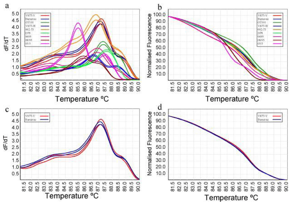
Infectious Bursal Disease virus (IBDVs) strain differentiation using HRM analysis: A TaqMan qRT-PCR and melting curve analysis could be used to trace mutations in the HVP2 region [15]. This method allowed comparing sequences between field and vaccinal strains [16,17]. TaqMan qRT-PCR was able to determine a single nucleotide polymorphism in VP2 [18]. Ghorashi, et al. [19] combined real-time RT-PCR and high resolution melt (HRM) curve analysis (Figure 1) to differentiate between classical vaccines/isolates and variant IBDV strains, which was developed into a robust technique for genotyping IBDV isolates/strains.
Nagoya Breed Differentiation using HRM assay: Native to Japan, the Nagoya breed of chicken is also known as the Nagoya Cochin. Given the seriousness of mislabelling chickens and the financial harm it does to the Nagoya breed’s production, a straightforward assay with high accuracy was needed to identify the Nagoya breed. Using a High-Resolution Melting (HRM) test, a unique screening assay was effectively developed to distinguish between the Nagoya breed. The ABR0417 microsatellite marker’s CA repeat was amplified using a primer set intended for HRM analysis. Using four differential chickens of the Nagoya breed and twelve other breeds, the applicability of the HRM-based technique was examined by comparing the results of the HRM analysis with those of the DNA sequence analysis. The melting curve and peak plots of the 16 chickens from the Nagoya breed were different from those of the other chickens, according to the HRM study of the birds. These findings showed that the HRM-based approach, which used the ABR0417 marker to identify the Nagoya breed, was a straightforward genetic test [20].
High-resolution melting analysis in the detection and discrimination of livestock infectious diseases
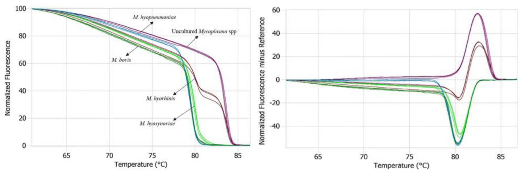
Mycoplasma phenotyping using HRM analysis: Numerous infectious diseases that affect both humans and animals are caused by Mycoplasma species. Ahani, et al. [21] evaluated the efficacy of a High-Resolution Melting curve assay (HRM) and real-time polymerase chain reaction (RT-PCR) for the quick identification of Mycoplasma species isolated from clinical cases of respiratory illness in cattle and pigs. Samples of lung from cows and pigs suspected of having respiratory infections were cultivated on a particular medium, and the extracted DNA was examined for Mycoplasma using standard polymerase chain reaction (PCR) techniques. For RT-PCR and HRM, a set of universal primers specific for the 16S ribosomal RNA gene was designed for five detection of distinct species of Mycoplasmas, including M. hyopneumoniae, M. bovis, M. hyorhinis, M. hyosynoviae, and additional uncultured Mycoplasma, could be distinguished using the HRM analysis. 16S rRNA gene sequencing was used to validate all findings. This quick and accurate test distinguished between bovine and porcine mycoplasmas at the species level, providing a straightforward substitute for PCR and sequencing (Figure 2) [22].
Mycoplasma mastitis is becoming more and more prevalent globally, and it has a big financial impact on the dairy sector. Early Mycoplasma mastitis diagnosis is crucial for disease management initiatives. Therefore, to screen for mycoplasma in dairy herds, a quick and precise diagnostic test is needed. Recently, High Resolution Melting curve analysis (HRM) was designed and is now frequently utilized for mycoplasma phenotyping at the strain or species level, among other organisms. This approach hasn’t been used to evaluate field isolates of mycoplasmas linked to mastitis or other milk environmental mollicutes. This study, conducted by Al-Farha, et al. [23] to develop a real-time, diagnostic polymerase chain reaction-High Resolution Melting curve analysis (PCR-HRM) method that would be appropriate for identifying and differentiating between five distinct mollicutes that were isolated at the cow level from a single commercial dairy farm in South Australia. An examination using real-time PCR-HRM analysis allowed for the identification and differentiation of five distinct mollicutes: A. laidlawii, M. arginini, M. bovirhinis, M. bovis, and uncultured Mycoplasma. Sequencing was used to validate the results. The test was designed to screen for Mycoplasma mastitis quickly and accurately.
High-resolution melting analysis in the detection and discrimination of companion animal infectious diseases
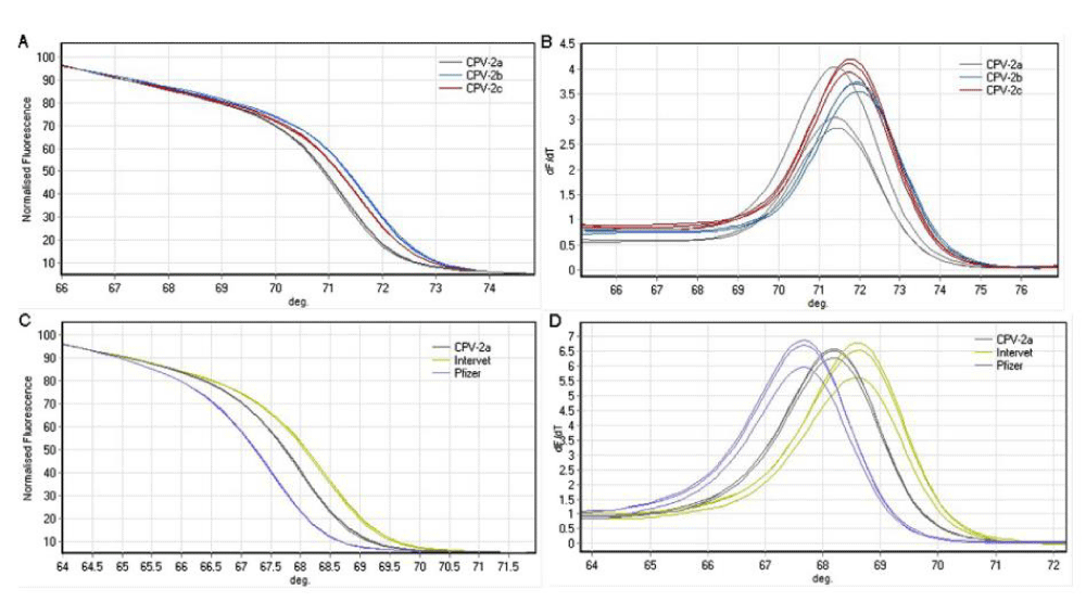
Bingga, et al. [24] developed a high-resolution melting (HRM) curve method to identify canine parvovirus type 2 (CPV2) strains by nested PCR. The viral VP2 capsid gene was used to create two sets of primers, CPV-426F/426R and CPV-87R/87F, which amplified a 52 bp and 53 bp product, respectively. The CPV-2a, CPV-2b, and CPV-2c A4062G and T4064A mutations were part of the region amplified by CPV-426F/ 426R. The A3045T mutation in the vaccination strains of CPV-2 and CPV-2a, CPV-2b, and CPV-2c was part of the region amplified by CPV-87F/87R. Using CPV-426F/426R, the PCR-HRM test was able to differentiate between CPV-2a, CPV-2b, and CPV-2c single nucleotide polymorphisms (Figure 3). The melting temperatures of CPV-2a, CPV-2b, and CPV-2c were different from one another (Table 1). After producing heteroduplexes using a CPV-2b reference sample, CPV-2b, and CPV-2c could be discriminated against based on the form of the melting curve. Using CPV-87F/87R, the vaccine strains of CPV-2 were recognized. This PCR-based HRM technique might be a good alternative to traditional, labor-intensive, expensive, or time-consuming CPV strain genotyping methods.
To detect and differentiate between Mycoplasma species that are often isolated from canine, avian, and ruminant samples, a real-time polymerase chain reaction assay called PCR-HRM was developed. One pair of universal primers specific for the spacer region between the 16S and 23S ribosomal RNA genes was utilized in the real-time PCR; a high-resolution melt fluorescent dye was employed to analyze the PCR product’s melting curve. The reference strains of M. arginini, M. bovigenitalium, M. bovis, M. bovirhinis, M. canadense, M. cynos, M. spumans, M. iowae, M. meleagridis, and M. agalactiae could all be distinguished using the real-time PCR-HRM assay. Testing field isolates of M. bovis, M. arginini, M. bovirhinis, M. bovigenitalium, M. iowae, and M. spumans allowed for the evaluation of the real-time PCR-HRM assay, and the results were in line with the fluorescent antibody test [25]. Real-time PCR-HRM was quicker and less expensive (1 - 2 hours) than the current gold standard IFAT and 16S rRNA gene sequence analysis, but it could only identify pure cultures. Although it could function on mixed-species colonies and takes around 6 hours to identify 6–10 isolates, the IFAT was technically challenging and prone to false positives due to autofluorescence and cross-reactions. The 16S rRNA gene could only be sequenced in pure cultures, was costly, and took two to three days to get results. The results of the investigation showed that the real-time PCR-HRM method was an efficient and quick assay for differentiating Mycoplasma species in pure cultures [25].
Canine Parvovirus (CPV) and Feline Parvovirus (FPV) are causative agents of diarrhea in dogs and cats, which in young animals shows up as diarrhea, vomiting, fever, appetite loss, leucopenia, and depression. Cats can become ill from single or combined CPV and FPV infections. An efficient virus diagnostic and genome typing technique with high sensitivity and specificity was needed to properly diagnose sick animals. Real-time PCR amplification was carried out by Sun, et al. [26], using a conserved region of the parvovirus that included one SNP, A4408C. Then, using Applied Biosystems® High-Resolution Melt Software v3.1, data were automatically analyzed and displayed. The outcomes demonstrated that both FPV and CPV could be found in the same PCR test. Canine adenovirus, canine coronavirus, and canine distemper virus did not cause any cross-reactions. The assay’s CPV and FPV genome copy count was limited to 4.2. The assay’s percentage of agreement with other methods was high. Thus, to accurately identify and differentiate between FPV and CPV in fecal samples, they created a diagnostic test that was also reasonably priced.
Infectious canine hepatitis (ICH) and Infectious Tracheobronchitis (ITB) are respectively caused by canine adenovirus types 1 (CAdV-1) and 2 (CAdV-2). A significant proportion of the canine population infected with CAdV-1 is asymptomatic, as evidenced by recent surveys and cases of ICH. An assay that is quick, highly sensitive, and specific was needed for the diagnosis of CAdV infection and differentiation between CAdV-1 and CAdV-2 because both CAdV types are detectable in the same biological matrices and viral coinfection with CAdV-1 and CAdV-2 is reported frequently. Melting curve analysis was used to optimize a SYBR Green real-time PCR assay to quickly differentiate between CAdV-1 and CAdV-2, and for detecting canine adenovirus in biological materials. The test that was designed demonstrated a high degree of sensitivity, repeatability, efficiency, and specificity in differentiating between the two kinds of CAdV. For simultaneous CAdV type identification, this dependable and quick method could offer a straightforward, practical, and affordable choice. It would be appealing to all diagnostic labs for both clinical and epidemiological research, and it would be practicable [27].
Babesia gibsoni and Babesia canis are the causative agents of canine babesiosis, a protozoal hemolytic illness. Serological testing and microscopic identification were used to diagnose the illness. To identify infections in dog breeds, a quantitative Fluorescence Resonance Energy Transfer (FRET)-PCR was developed. Melting curve analysis used the fluorescent probes’ disassociation temperature to distinguish between B. gibsoni, and B. canis canis/B. canis vogeli, and B. canis rossi. The occurrence of canine babesiosis is determined by environmental factors instead of hereditary predisposition, according to the study. B. gibsoni was eradicated by atovaquone and tilmicosin therapy, but not by doxcycline or berenil. This implied that real-time, high-resolution PCR analysis could enhance treatment monitoring and diagnosis [28].
Limitations of HRM analysis
HRM analysis is easy, quick, and affordable, but it is heavily dependent on high-quality PCR, tools and dyes. The type of single base substitution or the location of the variant within the PCR product are not relevant factors in heterozygote detection. It could be a little harder to find small insertions and deletions than substitutions. Critically, accuracy depends on the instrument’s resolution [29]. Instruments for high-resolution DNA melting can explain the discrepancies between expected and observed outcomes. The instrument employed had a significant impact on the apparent temperature variance within a genotype [30]. A review of this technology’s applicability for SSR genotyping by Yang, et al. [31] found significant drawbacks. These included the possibility of missing or incorrectly classifying genotypes when there were few alleles, as animals have a much smaller number of SSR loci than plants, making it difficult to select enough loci with low levels of polymorphism to be similar to those found in other species. The melting pattern may be impacted by the quality and quantity of DNA used in the experiment, which is another drawback of the HRM technique. Poor reproducibility was observed in the HRM curve analysis when amplicons that were not pre-amplified were used, either on different days or with different templates created from individual samples. The reference DNA templates, unknown samples acquired in the field, or DNA obtained using various extraction techniques must be pre-amplified by PCR for ensuing HRM analysis to get repeatable results using HRM [24]. It is expected that these restrictions will be overcome shortly as technology advances.
Future directions
Higher resolution and throughput could be possible with future advancements, opening up new applications. The efficiency of genetic analysis pipelines can be improved by integration with NGS procedures. HRM could be used often in clinical laboratories for diagnosis and mutation screening, as the technology develops and validation studies are carried out. HRM is going to be a big part of epigenetic research as it becomes more and more popular. With ongoing technical developments, broader applications in other domains, and integration with other molecular biology methods propelling its growth and significance, the future of HRM is bright.
Conclusion
HRM is a near-perfect assay for testing in the clinical laboratory, requiring less money and effort to design the assays. As discussed, HRM analysis is a practical analytical technique for identifying gene mutations in a wide range of pathogens. The constraint of HRM analysis for mutation and genotype screening is that it requires thorough validation studies before its implementation in clinical settings. Overall, HRM serves as a valuable tool in the veterinary arsenal, enhancing diagnostic accuracy, speeding up workflows, and contributing to improved animal health and welfare. All things considered, HRM has the potential to completely transform nucleic acid profiling in clinical and research contexts by getting beyond limitations about sensitivity and facilitating thorough examination of genetically varied materials. As a result of its influence on biomarker research and clinical microbiology, more precise and trustworthy diagnostic assays may be created, ultimately improving our understanding of disease mechanisms.
- Reed GH, Kent JO, Wittwer CT. High-resolution DNA melting analysis for simple and efficient molecular diagnostics. Pharmacogenomics. 2007 Jun;8(6):597-608. doi: 10.2217/14622416.8.6.597. PMID: 17559349.
- Chatzidimopoulos M, Ganopoulos I, Moraitou-Daponta E, Lioliopoulou F, Ntantali O, Panagiotaki P, Vellios EK. High-resolution melting (HRM) analysis reveals genotypic differentiation of Venturia inaequalis populations in Greece. Frontiers in Ecology and Evolution. 2019; 7: 489.
- Chatzidimopoulos M, Ganopoulos I, Madesis P, Vellios E, Tsaftaris A, Pappas AC. High‐resolution melting analysis for rapid detection and characterization of Botrytis cinerea phenotypes resistant to fenhexamid and boscalid. Plant pathology. 2014; 63 (6):1336-1343.
- Słomka M, Sobalska-Kwapis M, Wachulec M, Bartosz G, Strapagiel D. High Resolution Melting (HRM) for High-Throughput Genotyping-Limitations and Caveats in Practical Case Studies. Int J Mol Sci. 2017 Nov 3;18(11):2316. doi: 10.3390/ijms18112316. PMID: 29099791; PMCID: PMC5713285.
- Machuve D, Nwankwo E, Mduma N, Mbelwa J. Poultry diseases diagnostics models using deep learning. Front Artif Intell. 2022; 5:733345.
- Alghizzawi D, Ababneh MM, Al-Zghoul MB. Differentiation of avian orthoavulavirus 1, genotype VII and its sub-genotypes by high resolution melting (HRM) assay. Pak Vet J. 2024; 44(1):47-54.
- Dimitrov KM, Ramey AM, Qiu X, Bahl J, Afonso CL. Temporal, geographic, and host distribution of avian paramyxovirus 1 (Newcastle disease virus). Infect Genet Evol. 2016 Apr;39:22-34. doi: 10.1016/j.meegid.2016.01.008. Epub 2016 Jan 12. PMID: 26792710.
- Li Z, Wang X, Zhang R, Chen J, Xia L, Lin S, Xie Z, Jiang S. Evidence of possible vertical transmission of duck circovirus. Vet Microbiol. 2014 Nov 7;174(1-2):229-32. doi: 10.1016/j.vetmic.2014.09.001. Epub 2014 Sep 16. PMID: 25263494.
- Wan C, Cheng L, Fu G, Shi S, Chen H, Fu Q, Liu R, Huang Y. Establishment a PCR-RFLP method for rapid differential duck circovirus genotype 1 and genotype 2. Chin Virol Poult. 2016;38:53-55.
- Fu H, Zhao M, Chen S, Huang Y, Wan C. Simultaneous detection and differentiation of DuCV-1 and DuCV-2 by high-resolution melting analysis. Poult Sci. 2024 Apr;103(4):103566. doi: 10.1016/j.psj.2024.103566. Epub 2024 Feb 15. PMID: 38417341; PMCID: PMC10907865.
- Ababneh M, Ababneh O, Al-Zghoul MB. High-resolution melting curve analysis for infectious bronchitis virus strain differentiation. Vet World. 2020 Mar;13(3):400-406. doi: 10.14202/vetworld.2020.400-406. Epub 2020 Mar 3. PMID: 32367941; PMCID: PMC7183480.
- Kirkpatrick NC, Blacker HP, Woods WG, Gasser RB, Noormohammadi AH. A polymerase chain reaction-coupled high-resolution melting curve analytical approach for the monitoring of monospecificity of avian Eimeria species. Avian Pathol. 2009 Feb;38(1):13-9. doi: 10.1080/03079450802596053. PMID: 19156576.
- Steer PA, O'Rourke D, Ghorashi SA, Noormohammadi AH. Application of high-resolution melting curve analysis for typing of fowl adenoviruses in field cases of inclusion body hepatitis. Aust Vet J. 2011 May;89(5):184-92. doi: 10.1111/j.1751-0813.2011.00695.x. PMID: 21495991.
- Chamings A, Hewson KA, O'Rourke D, Ignjatovic J, Noormohammadi AH. High-resolution melt curve analysis to confirm the presence of co-circulating isolates of avian nephritis virus in commercial chicken flocks. Avian Pathol. 2015;44(6):443-51. doi: 10.1080/03079457.2015.1085648. PMID: 26365395.
- Jackwood DJ, Spalding BD, Sommer SE. Real-time reverse transcriptase-polymerase chain reaction detection and analysis of nucleotide sequences coding for a neutralizing epitope on infectious bursal disease viruses. Avian Dis. 2003 Jul-Sep;47(3):738-44. doi: 10.1637/6092. PMID: 14562905.
- Jackwood DJ, Sommer SE. Virulent vaccine strains of infectious bursal disease virus not distinguishable from wild-type viruses with marker the use of a molecular. Avian Dis. 2002 Oct-Dec;46(4):1030-2. doi: 10.1637/0005-2086(2002)046[1030:VVSOIB]2.0.CO;2. PMID: 12495070.
- Gao HL, Wang XM, Gao YL, Fu CY. Direct evidence of reassortment and mutant spectrum analysis of a very virulent infectious bursal disease virus. Avian Dis. 2007 Dec;51(4):893-9. doi: 10.1637/7626-042706R1.1. PMID: 18251399.
- Wu CC, Rubinelli P, Lin TL. Molecular detection and differentiation of infectious bursal disease virus. Avian Dis. 2007 Jun;51(2):515-26. doi: 10.1637/0005-2086(2007)51[515:MDADOI]2.0.CO;2. PMID: 17626477.
- Ghorashi SA, O'Rourke D, Ignjatovic J, Noormohammadi AH. Differentiation of infectious bursal disease virus strains using real-time RT-PCR and high resolution melt curve analysis. J Virol Methods. 2011 Jan;171(1):264-71. doi: 10.1016/j.jviromet.2010.11.013. Epub 2010 Nov 24. PMID: 21111004.
- Aoki A, Mori Y, Okamoto Y, Jinno H. A high-resolution melting-based assay for discriminating a native Japanese chicken, the Nagoya breed, using the ABR0417 microsatellite marker. Eur Food Res Technol. 2024;250(3):745-750.
- Ahani Azari A, Amanollahi R, Jafari Jozani R, Trott DJ, Hemmatzadeh F. High-resolution melting curve analysis: a novel method for identification of Mycoplasma species isolated from clinical cases of bovine and porcine respiratory disease. Trop Anim Health Prod. 2020 May;52(3):1043-1047. doi: 10.1007/s11250-019-02098-4. Epub 2019 Nov 1. PMID: 31673887; PMCID: PMC7222993.
- Fraley SI, Hardick J, Masek BJ, Athamanolap P, Rothman RE, Gaydos CA, Carroll KC, Wakefield T, Wang TH, Yang S. Universal digital high-resolution melt: a novel approach to broad-based profiling of heterogeneous biological samples. Nucleic Acids Res. 2013 Oct;41(18):e175. doi: 10.1093/nar/gkt684. Epub 2013 Aug 9. Erratum in: Nucleic Acids Res. 2016 Jan 8;44(1):508. Jo Masek, Billie [Corrected to Masek, Billie J]. PMID: 23935121; PMCID: PMC3794612.
- Al-Farha AA, Petrovski K, Jozani R, Hoare A, Hemmatzadeh F. Discrimination between some Mycoplasma spp. and Acholeplasma laidlawii in bovine milk using high resolution melting curve analysis. BMC Res Notes. 2018 Feb 7;11(1):107. doi: 10.1186/s13104-018-3223-y. PMID: 29415764; PMCID: PMC5804061.
- Bingga G, Liu Z, Zhang J, Zhu Y, Lin L, Ding S, Guo P. High resolution melting curve analysis as a new tool for rapid identification of canine parvovirus type 2 strains. Mol Cell Probes. 2014 Oct-Dec;28(5-6):271-8. doi: 10.1016/j.mcp.2014.08.001. Epub 2014 Aug 23. PMID: 25159576.
- Rebelo AR, Parker L, Cai HY. Use of high-resolution melting curve analysis to identify Mycoplasma species commonly isolated from ruminant, avian, and canine samples. J Vet Diagn Invest. 2011 Sep;23(5):932-6. doi: 10.1177/1040638711416846. Epub 2011 Aug 19. PMID: 21908349.
- Sun Y, Cheng Y, Lin P, Zhang H, Yi L, Tong M, Cao Z, Li S, Cheng S, Wang J. Simultaneous detection and differentiation of canine parvovirus and feline parvovirus by high resolution melting analysis. BMC Vet Res. 2019 May 10;15(1):141. doi: 10.1186/s12917-019-1898-5. PMID: 31077252; PMCID: PMC6511188.
- Balboni A, Dondi F, Prosperi S, Battilani M. Development of a SYBR Green real-time PCR assay with melting curve analysis for simultaneous detection and differentiation of canine adenovirus type 1 and type 2. J Virol Methods. 2015 Sep 15;222:34-40. doi: 10.1016/j.jviromet.2015.05.009. Epub 2015 May 29. PMID: 26028428.
- Wang C, Ahluwalia SK, Li Y, Gao D, Poudel A, Chowdhury E, Boudreaux MK, Kaltenboeck B. Frequency and therapy monitoring of canine Babesia spp. infection by high-resolution melting curve quantitative FRET-PCR. Vet Parasitol. 2010 Feb 26;168(1-2):11-8. doi: 10.1016/j.vetpar.2009.10.015. Epub 2009 Oct 28. PMID: 19931290.
- Wittwer CT. High-resolution DNA melting analysis: advancements and limitations. Hum Mutat. 2009 Jun;30(6):857-9. doi: 10.1002/humu.20951. PMID: 19479960.
- Herrmann MG, Durtschi JD, Wittwer CT, Voelkerding KV. Expanded instrument comparison of amplicon DNA melting analysis for mutation scanning and genotyping. Clin Chem. 2007 Aug;53(8):1544-8. doi: 10.1373/clinchem.2007.088120. Epub 2007 Jun 7. PMID: 17556647.
- Yang M, Yue YJ, Guo TT, Han JL, Liu JB, Guo J, Sun XP, Feng RL, Wu YY, Wang CF, Wang LP, Yang BH. Limitation of high-resolution melting curve analysis for genotyping simple sequence repeats in sheep. Genet Mol Res. 2014 Apr 8;13(2):2645-53. doi: 10.4238/2014.April.8.7. PMID: 24782053.
Article Alerts
Subscribe to our articles alerts and stay tuned.
 This work is licensed under a Creative Commons Attribution 4.0 International License.


 Save to Mendeley
