Babesia Microti – Known and Unknown Protists
1Department of Skin Structural Studies, School of Pharmacy with the Division of Laboratory Medicine in Sosnowiec, Medical University of Silesia in Katowice, Poland
2Department of Animal Histology and Embryology, Faculty of Biology and Environmental Protection, University of Silesia in Katowice, Poland
3Institute of Materials Science, Faculty of Computer Science and Materials Science, University of Silesia in Katowice, Poland
Author and article information
Cite this as
Albertyńska M, Rupik W, Hermyt M, Okła H, Jasik KP (2017) Babesia Microti – Known and Unknown Protists. Glob J Zool. 2017; 2(1): 001-007. Available from: 10.17352/gjz.000004
Copyright License
© 2017 Albertyńska M. This is an open-access article distributed under the terms of the Creative Commons Attribution License, which permits unrestricted use, distribution, and reproduction in any medium, provided the original author and source are credited.B. microti is known as the main etiological agent of human babesiosis and there are some case studies for that disease, highlighting the fact that this is an important “emerging tick-borne disease”. However a lot of information about this protist is unclear.
The reactions in the liver are noticeable already after the fi rst three weeks of infection. This therefore provides the basis for discussion of the effect of B. microti contact with hepatocytes in vitro and in vivo. This issue is an essential objective of our study.
The effect of B. microti on the liver as an organ and the individual hepatocytes is evident. This may be an indirect impact, through the various factors of metabolic pathways. Ultrastructural studies of breeding co-culture of hepatocytes and B. microti has shown, however, that piroplasms B. microti are adhering to the outer surface of the hepatocytes. This contact is associated with destructive changes in hepatocytes both necrotic and apoptotic.
Babesia microti is a species of the phylum Apicomplexa (Piroplasmida) and invades red blood cells of vertebrates. B. microti is known to be a cosmopolitan species, but the main areas of its occurrence are the United States and European countries such as: France, England, Ireland, Germany, Spain, Switzerland, Russia and Poland [1,2]. In the US, these piroplasm have been reported mainly in the northeastern and midwestern States [3,4].
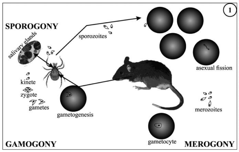
The life cycle of B. microti is associated with the metagenesis and runs with two hosts: intermediate – vertebrate, and the definitive - the tick. Protozoan parasites reproduce asexually in the intermediate hosts, while sexual reproduction occurs in the definitive host [5-7].
The main intermediate hosts and reservoirs of these protists are rodents (Apodemus flavicolii, Clethrionmys glaveolus, Sorex araneas). B. microti may be optionally prevalent human parasite.
Typical final hosts and vectors of B. microti in Europe are Ixodes ricinus and I. trianguliceps [8], and in North America - I. scapularis [9].
Despite B. microti is known as the main etiological factor of the human babesiosis and there are the case reports pertaining to this disease which indicated the fact that it is “Emerging tick-borne disease” [10] there are still many inconsistencies and unsolved issues. This issues are associated with the taxonomy of this pathogen, its pathogenesis and the correlation between the pathogen and the host. Commonly used name of B. microti is often negated and these piroplasms alternatively are called Theileria microti [11]. This is explained by the fact that in the course of the asexual reproduction in the intermediate host, piroplasms invade not only erythrocytes but also the lymphocytes, which is contrary to members of genus Babesia [11].
However the genome of B. microti has been sequenced. These analysis shows that this species does not belong to none of the established genera - Babesia and Theileria - but to a separate genus [12].
A typical life cycle of Babesia (Theileria) microti presented in the literature [13] includes merogony and forming the pre-gametocytes in intermediate hosts, and gamogony and sporogony in definitive hosts (Figure 1).
Babesiosis was first described in 1888 by Victor Babeş, in cattle with symptoms of fever and haemoglobinuria. After 9 years, it was found that the etiological agent of this disease is transmitted by ticks. Human babesiosis was first found in 1957 on the site of the former Yugoslavia. In the United States, the first case of human piroplasmosis has been described in 1966, while in Poland in 1997 [14-16].
Babesiosis is known to be endemic disease widespread in temperate, tropical and subtropical climate. In a temperate climate babesiosis occurs in Europe and North America. In Europe, babesiosis mainly occurs in France, England, Ireland, Germany, Spain, Switzerland, Russia, Poland and in the former Yugoslavia. While in the United States the main areas of the prevalence of human babesiosis are northeastern and midwestern States.
In tropical and subtropical climate babesiosis occurs in Africa, Asia (China, Taiwan, and India) and Australia [16].
Clinical signs of human babesiosis appear between 1 and 4 weeks from man’s prick by a tick infected with B. microti or between 9 weeks and 6 months in the case of babesiosis after blood transfusion.
Sporozoites penetrating into the interior of the cell cause the physical, chemical and structural changes of cell membrane.
Inside the erythrocytes sporozoites are divided into 2-4 merozoites, and formed in this way merozoites leave the red blood cell, causing its damage and degradation.
The effects of merogony are hemolytic anemia, thrombocytopenia, increased number of reticulocytes and low hematocrit.
Physical and chemical changes of membranes of vascular endothelial cells, thrombocytes and red blood cells are associated with occlusion of the small blood vessels and the formation of blockages. In the course of babesiosis, the characteristic signs include splenomegaly, hepatomegaly and dysfunction of the liver, kidneys and central nervous system [6,16,17].
During the course of the disease activation of the complement system and production of pro-inflammatory cytokines occurs, which results in emerging flu-like symptoms. It also causes stimulatation of suppressor T cells (stimulation of primary) and B cells (stimulating secondary). Blood chemistry shows increase in liver enzymes, such as transaminases, alkaline phosphatase, lactate dehydrogenase and free bilirubin [17].
The clinical course babesiosis in humans may be asymptomatic, mild or severe. Statistically at 25% of adults and 50% of children, babesiosis occurs asymptomatic and sometimes it ends with a self-healing or goes into latent phase [18].
However, babesiosis can be deadly, especially for the elderly or immunocompromised [18,19].
B. microti as a representative of the Apicomplexa is usually compared with Theileria spp. and Plasmodium spp. The effect of these comparisons draws attention to the lack of tropism Babesia spp. to lymphocytes, in contrast to Theileria spp. and the inability to penetrate piroplasm of the genus Babesia to hepatocytes, in contrast to the representatives of the Plasmodium spp., which in the first period of invasion penetrate to the liver cells. However, analysing of the etiology of babesiosis in rats draws attention to the fact that piroplasms induce significant changes in hepatocytes [20]. The first reactions in the liver are noticeable already after the first three weeks of infection. This therefore provides the basis for discussion of the effect of B. microti contact with hepatocytes in vitro and in vivo. This issue is an essential objective of the study.
Material and Methods
The research was conducted in both in vivo and in vitro.
in vivo analyses (light microscopy observations)
The animals were earlier infected by reference strain culture of B. microti (ATCC 30221). Infection was caused by intraperitoneal (i.p.) injection of 0.5 mL protozoan culture in the six-week-old rats. The animals were housed in standardized conditions: temperature aprox. 20-22oC, humidity – 50-60% and lighting cycles – 12 hours of light and 12 hours of darkness. The breeding was conducted in cages with pine sawdust which was autoclaved. The animals had unlimited access to water and fodder. Control material was livers collected from 2 healthy rats. During the experimental steps, 0.5 mL sodium chloride physiological solution for intravenous injection (Inj. Natrii Chlorati Isotonica Polpharma, 9 mg/mL; Polpharma, Starogard Gdanski, Poland) was injected intraperitoneal in the rats. Control rats were housed in same conditions as animals infected with B. microti.
Detection of parasitemia in blood smears was performed using commercially available Histology FISH Accessory Kit (see below).
Three week after inoculation, the animals were euthanized by inhalation anesthesia with isoflurane (Forane® - Baxter, Deerfield, IL, USA). During post-mortem examinations, liver samples were collected for histological analysis.
Detection of B. microti genetic material in liver specimens and blood smears was performed using commercially available Histology FISH Accessory Kit (DAKO, Carpinteria, CA, USA). The preparation procedures of tissues’ slides and smears of blood were in step with the manufacturer’s instructions. In this study, the fluorescein-labeled probe complementary to the fragment of B. microti 18S rDNA gene was used. The probe sequence was as follows: 5’-fluorescein-GCCACGCGAAAACGCGCCTCGA-fluorescein-3. The oligonucleotide synthesis was performed in the Institute of Biochemistry and Biophysics in Warsaw.
After the hybridization step, the rinses were conducted according to the manufacturer’s instructions. The analysis of preparations was performed with Olympus BX60 epifluorescent microscope with 150W xenon lamp using blue filter for fluorescein.
Tissues were fixed in Bouin’s solution at room temperature. In the next stage tissues were rinsed in 80% ethyl alcohol and then embedded in paraffin blocks according to standard histological procedure. Paraffin sections (6-µm thick) were stained by the Masson trichrome method. The dried preparations were mounted in synthetic resin DPX Mounting Medium (POCh, Gliwice, Poland) and analyzed using Olympus BX60 microscope equipped with XC50 digital camera and Olympus cellSens Standard software.
The animal use in this study was approved by the Local Ethics Committee for Animal Experiments in Katowice (Resolution number: 32/2011 dated May 23, 2011).
In vitro analyses (light and transmission electron microscopy observations)
In this study AML 12 line of normal hepatocytes (ATCC CRL-2254) was infected by reference strain culture of B. microti (ATCC 30221).
Two samples: infected and uninfected hepatocytes were cultured in DMEM: F-12 Medium (ATCC, London, UK) with 0.005 mg/ml insulin, 0.005 mg/ml transferrin, 5 ng/ml selenium, 40 ng/ml dexamethasone and 10% fetal bovine serum, in culture flasks (Nunclon ™ Sphera ™ Flasks, culture area: 25 cm2) with bacteriological filters (Nunc, Wiesbaden, Germany). The culture conditions were temperature 37oC, 5% CO2. Infected cultures of hepatocytes cell line and the control were incubated for 72 hours.
After this time, the hepatocytes cultures were fixed in a solution of 2.5% paraformadehyde in phosphate buffer at 4°C. After washing with 0.1 M phosphate buffer (pH 7.4), the tissues were postfixed in buffered 1% osmium tetroxide solution (Sigma-Aldrich, St. Louis, MO, USA) for 2 hours. In the next step the samples were rinsed with phosphate buffer, dehydrated in a series of ethyl alcohol and acetone according to standard histological procedures. Dehydrated tissue was embedded in epoxy resin-Poly/Bed 812® media Embeeding/DMP-30 Kit (Polyscience, Inc., Warrington, PA, USA). The polymerization of the resin was carried out at 60°C for 72 hours. All procedures were performed in culture flasks.
Semi-thin sections (0.5 µm thick) were stained with methylene blue (AppliedChem, Darmstadt, Germany) and observed under a light microscope. In contrast, ultra-thin sections (80 nm thick) were stained with uranyl acetate (Polyscience, Inc., Warrington, PA, USA) and lead citrate (Sigma-Aldrich, St. Louis, MO, USA).
The ultrastructural observations were conducted with Hitachi H500 (Hitachi Ltd, Tokyo, Japan) transmission electron microscope at an acceleration voltage of 75 kV.
Additionally some cultures of infected and uninfected hepatocytes were incubated in 1% sodium citrate for 2 minutes at 4°C. Slides were washed in TBS (Sigma-Aldrich. St. Louis, MO, USA) three times for 5 minutes and stained with a TUNEL (Tdt-mediated deoxyUridine triphosphate-biotin/digoxigenin Nick End-Labeling) reaction mixture for 1 hour at 37°C in the dark. All steps were performed according to manufacturer’s protocol (In situ cell Death Detection Kit, TRM red; Roche, Mannheim, Germany). Furthermore all of the slides with cell cultures were stained with DAPI method. The material was analyzed using an Olympus BX60 fluorescent microscope (Olympus Corp., Tokyo, Japan).
Results
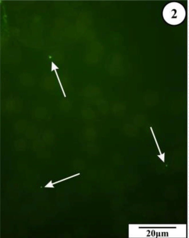
The presence of B. microti DNA was demonstrated in blood cells using FISH method (Figure 2).
The spot signals of fluorescence were also observed in the samples of infected rat’s liver (Figure 3).
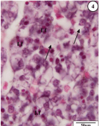
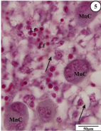
After 3 weeks of invasion by Babesia microti, observation of rats liver microscopic sections shows that between the hepatocytes there is a large amounts of big, polynuclear cells - macrophages (Figure 4). Perivascular lymphocyte infiltrations are visible (Figure 5). Additionally, in the course of babesiosis there were also observed numerous hepatocyte damages. The cells were swollen with the forming vacuoles in the cytoplasm (Figure 4).
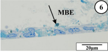
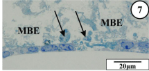
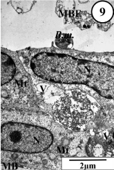
Light microscopic and ultrastructural analyses of the hepatocytes culture has shown the contact between the hepatocytes and piroplasms B. microti. The presence of piroplasms inside cultured cells is ambiguous and questionable. Above the free surface of hepatocytes monolayer there are many leftover membranes after erythrocytes from the inoculum. In some places, oval structures containing the cell nucleus with a diameter of approx. 1-3 µm, adhere to the outer membrane of cultured hepatocytes (Figure 6). In the cross sections by a layer of infected hepatocytes there are visible also the places in which the layer is discontinuous and the cells in these places are destroyed. Images of the destruction of cells are heterogeneous, as they can be subtle and the small fragments of individual cells that have been disintegrated - apoptotic bodies - can be noticed (Figure 7). In others fields of observation there are large spaces with vacuolization of fragments of the cells. In the latter case, the degenerative changes are more extensive (Figure 7).
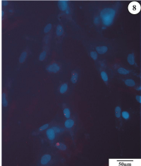
The TUNEL reaction showed a lot of fluorescent signals. The apoptotic cell death was detected in the hepatocytes infected with B. microti (Figure 8)
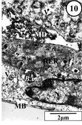
Analogous phenomenon of piroplasm adhesion to the free surface of hepatocytes in culture in vitro has been noticed in the ultrastructural observations in the TEM. In the ultrathin sections of some hepatocytes there appear swollen mitochondria with osmiophobic matrix, and a large vacuoles with granular-filamentous material. Such material also occurs in the extracellular spaces between hepatocytes (Figures 9,10).
Discussion
According to the literature Babesia microti penetrates directly into erythrocytes without the stages of initial proliferation at other organs [5,21].This feature is related to the specificity of the impact of the membrane of red blood cells with a particular species of piroplasm [22,23].
The most common complications of severe babesiosis include acute respiratory failure, congestive heart failure, liver and renal failure and splenic rupture [24,25].
Impact of B. microti in different organs is complicated and there are later effects of the sequestration and of capillaries’ thromboembolism.
Different analyses indicated changes in hepatic enzymes in the blood serum. In the course of babesiosis in humans and animals an increase in aspartate aminotransferase and alanine aminotransferase activities was observed [24,26,27].
In rats infected with B. microti notes lymphocytic infiltrations, an increased number of large polynuclear cells, as well as a significant vacuolization changes in hepatocytes. Noticeable is also the correlation of various occurrence. Groups of lymphocytes are a symptom of the immune response, while polynuclear, large cells may be associated with the processes of regeneration or forming macrophages [28]. These cells serve an important role in the final stages of apoptotic changes.
Vacuolization of hepatocytes indicates clearly the advancing of destructive processes of necrosis. The biochemical analyses detected that babesiosis causes decrease the activity of antioxidant system, reflected by reduced glutathione level and catalase activity, and total antioxidant capacity of the of liver. Therefore hepatic artery embolization in tissue is more sensitive to damaging factors [29-31].
In vitro observations allowed to draw attention to the fact that although there have not been shown unequivocally that B. microti penetrates hepatocytes, however it was stated that piroplasms released from erythrocytes adhere to the hepatocytes plasmalemma. The molecular mechanism of the contact between hepatocytes and B. microti require further study, however, the analysis of the ultrastructure confirms the observations based on routine histological analyses and allows to notice the destructive effect of parasites on the liver cells. Characteristic is the mitochondrial damage and the cells’ vacuolization. These changes indicate the initial stage of necrosis. It can therefore be concluded that the B. microti in infected rats causes both necrotic and apoptotic changes.
Our studies confirm the previously described hepatic tissue changes in rats infected long-term with Babesia microti [20].
The intense apoptotic processes in hepatocytes cultured with B. microti we confirmed with use of the TUNEL method, however, the mechanism of induction of this phenomenon requires further analysis.
The interesting thing is that the processes of apoptosis in hepatocytes are clearly hampered by Plasmodium spp. However at late stages, upon completion of schizogony, a novel form of host cell death is triggered. It displays some hallmarks of apoptosis, but also marked differences, such as the failure of the infected hepatocytes to activate caspases and to display phosphatidylserine on its surface of the host cell [32].
The influence of protozoan parasites on death of the host cells has been shown in many publications. It was noted that the impact can be differentiated both in terms of stage on which it occurs as well as the final effect, i.e., whether the inhibition or stimulation of the cell death process takes place [33,34]. However it is characteristic, that the modulation of the cell death process involves mainly cells that are engaged in the life cycle of the given species [34].
Thus remains unanswered question, what is the meaning of hepatocytes for the Babesia microti development.
Summary
Babesia microti is an intraerythrocytic parasite, however, the impact of this species on the host organism is multilateral. Although it is not clearly demonstrated the ability to infect by B. microti other cells than erythrocytes and lymphocytes, possibly, however, the impact of these piroplasm in different organs is very strong and diverse.
The effect of B. microti on the liver as an organ and the individual hepatocytes is evident. This may be an indirect impact, through the various factors of metabolic pathways. Ultrastructural studies of breeding co-culture of hepatocytes and B. microti has shown, however, that piroplasms B. microti are adhere to the outer surface of the hepatocytes. This contact is associated with destructive changes in hepatocytes both necrotic and apoptotic.
This study was supported by the grant No KNW-1-039/N/5/0 from Medical University of Silesia in Katowice, Poland. The authors are deeply indebted to Michelle L. Simmons from University of Silesia, Katowice, Poland for improving the English.
- Berger S (2016) Babesiosis: Global Status In GIDEON Informatics, Los Angeles, USA 9-25. Link: https://goo.gl/KWP9JY
- Vannier E, Krause PJ (2012) Human babesiosis. N Engl J Med 366: 2397-2407. Link: https://goo.gl/prh8hr
- Diuk-Wasser MA, Liu Y, Steeves TK, Folsom-O’Keefe C, Dardick KR, et al. (2014) Monitoring Human Babesiosis Emergence through Vector Surveillance New England. Emerg Infect Dis 20: 225-231. Link: https://goo.gl/Z3piUy
- Vannier E, Krause PJ (2009) Update on babesiosis. Interdisciplinary Perspectives on Infectious Diseases. 9: 9 Link: https://goo.gl/5O30Ev
- Sonenshine DE (2014) Tick-borne Protozoa Chapter 6 In: Biology of Ticks, Oxford University Press: New York, USA. 147-179 Link: https://goo.gl/BXfDKz
- Homer MJ, Aguilar-Delfin I, Telford SR III, Krause PJ, Persing DH (2000) Babesiosis. Clinical Microbiology Reviews 13: 451-469. Link: https://goo.gl/FDPN2D
- Rożej-Bielicka W, Stypułkowska-MisiurewiczH, GołąbE (2015)Human babesiosis. Przegląd Epidemiologiczny 69: 489–494. Link: https://goo.gl/68A4Up
- Capligina V, Berzina I, Bormane A, Salmane I, Vilks K, et al. (2016) Prevalence and phylogenetic analysis of Babesia spp. in Ixodes ricinus and Ixodes persulcatus ticks in Latvia. Exp Appl Acarol 68: 325–336. Link: https://goo.gl/FVe9lk
- Diuk-Wasser M, Vourc’h G, Cislo P, Gatewood Hoen A, Melton F, et al. (2010) Field and climate-based model for predicting the density of host-seeking nymphal Ixodes scapularis , an important vector of tick-borne disease agents in the eastern United States. Global Ecology and Biogeography 19: 504–514. Link: https://goo.gl/L40tXf
- Smith R, Elias SP, Borelli TJ, Missaghi B, York BJ, et al. (2014) Human Babesiosis, Maine, USA, 1995–2011. Emerging Infectious Diseases 20: 1727-1730. Link: https://goo.gl/IqW83c
- Chiodini PL (2003) Babesiosis. In eds. Manson's tropical diseases. 21st edition. Edited by Cook GC, Zumla AI, Saunders, London 1297–1301. Link:
- Cornillot E, Hadj-Kaddour K, Dassouli A, Noel B, Ranwez V, et al. (2012)Sequencing of the smallest Apicomplexan genome from the human pathogen Babesia microti. Nucleic Acids Research40: 9102–9114. Link: https://goo.gl/RPibAx
- Chauvin A, Moreau E, Bonnet S, Plantard O, Malandrin L (2009) Babesia and its hosts: adaptation to long-lasting interactions as a way to achieve efficient transmission. Vet Res 40: 37. Link: https://goo.gl/MbqKyY
- Hunfeld KP, Hildebrandt A, Gray JS (2008) Babesiosis: recent insights into an ancient disease. Int J Parasitol 38: 1219-1237. Link: https://goo.gl/4XrWTy
- Hildebrandt A, Gray JS, Hunfeld KP (2013) Human babesiosis in Europe: what clinicians need to know. Infection 41: 1057-1072. Link: https://goo.gl/BNYWoQ
- Vannier E, Krause PJ (2012) Human babesiosis. New Engl J Med 366: 2397–2407. Link: https://goo.gl/MS43nS
- Vannier E, Gewurz BE, Krause PJ (2008) Human babesiosis. Infectious Disease Clinics of North America 22: 469-488. Link: https://goo.gl/quQTvx
- Krause PJ, Daily J, Telford SR, Vannier E, Lantos P, et al. (2007) A. Shared features in the pathobiology of babesiosis and malaria. Trends in Parasitology 23:605-610. Link: https://goo.gl/UZQYWy
- Joseph JT, Roy SS, Shams N, Visintainer P, Nadelman RB, et al. (2011) Babesiosis in Lower Hudson Valley, New York, USA. Emerging Infectious Diseases 17: 843-847 Link: https://goo.gl/w9GcpG
- Okła H, Jasik KP, Słodki J, Rozwadowska B, Słodki A, et al. (2014) Hepatic Tissue Changes in Rats Due to Chronic Invasion of Babesia microti . Folia Biologica 62: 353-359. Link: https://goo.gl/6GqBqL
- Borggraefe I, Yuan J, Telford SR III, Menon S, Hunter R, et al. (2006) Babesia microti primarily invades mature erythrocytes in mice. Infection and Immunity 74: 3204–3212. Link: https://goo.gl/ZVZw7N
- Park H, Hong SH, Kim K, Cho SH, Lee WJ, et al. (2015)Characterizations of individual mouse red blood cells parasitized by Babesia microti using 3-D holographic microscopy. Scientific Reports 5: Article number: 10827. Link: https://goo.gl/Usn1wm
- Heussler VT, Machado J Jr, Fernandez PC, Botteron C, Chen CG, et al. (1999) The intracellular parasite Theileria parva protects infected T cells from apoptosis, Proceedings of the National Academy of Sciences 96: 7312–7317. Link: https://goo.gl/lXbE2m
- Hatcher JC, Greenberg PD, Antique J, Jimenez- Lucho VE (2001) Severe babesiosis in Long Island: review of 34 cases and their complications. Clinical Infectious Diseases 32: 1117-1125. Link: https://goo.gl/qMcWvE
- Zhou X, Xia S, Huang JL, Tambo E, Zhuge HX, et al. (2014) Human babesiosis, an emerging tick-borne disease in the People’s Republic of China. Parasites & Vectors 7: 509. Link: https://goo.gl/PEVyK4
- Barić Rafaj R, Mrljak V, Kučer N, Brkljačić M, Matijatko V (2007) Protein C activity in babesiosis of dogs. Veterinarski arhiv 77: 1-8. Link: https://goo.gl/h4uvIk
- Dkhil MA, Al-Quraishy S, Abdel-Baki AS (2010) Hepatic tissue damage induced in Meriones ungliculatus due to infection with Babesia divergens -infected erythrocytes. Saudi Journal of Biological Sciences 17: 129-132. Link: https://goo.gl/oq6Dw7
- Zhou X, Platt JL (2011) Molecular and Cellular Mechanisms of Mammalian Cell Fusion. Chapter 4, pp 33-64 In: Link: https://goo.gl/Yzjsca
- Dittmar T, Zänker KS (2011) Advances in Experimental Medicine and Biology: I Cell Fusion in Health and Disease. Springer Science+Business Media B.V. Link: https://goo.gl/bgkleb
- Dhkil MA, Abdel-Baki AS, Al-Quraishy S, Abdel-Moneim AE (2013) Hepatic oxidative stress in Mongolian gerbils experimentally infected with Babesia divergens . Ticks and Tick Borne Disease 4: 346-451. Link: https://goo.gl/LqZKgF
- El-Far AH, Bakeir NA, Shaheen HM (2014) Anti-Oxidant Status for the Oxidative Stress in Blood of Babesia Infested Buffaloes. Global Veterinaria 12: 517-522. Link: https://goo.gl/T5tgTu
- Hamid OMA, Radwan MEI, Ali AF (2014) Biochemical changes associated with babesiosis infested cattle. Journal of Applied Chemistry 7 : 87-92. Link: https://goo.gl/DhqiZX
- Heussler VT, Stanway RR (2008) Cellular and molecular interactions between the apicomplexan parasites plasmodium and Theileria and their host cells. Parasite 15: 211-218. Link: https://goo.gl/w9cKU1
- Lüder CG, Stanway RR, Chaussepied M, Langsley G, Heussler VT (2009) Intracellular survival of apicomplexan parasites and host cell modification. Int J Parasitol 39: 163-173. Link: https://goo.gl/2IZUdW
- Carmen JC, Sinai AP (2007) Suicide prevention: disruption of apoptotic pathways by protozoan parasites. Molecular Microbiology 64: 904-916. Link: https://goo.gl/4T1YHl
Article Alerts
Subscribe to our articles alerts and stay tuned.
 This work is licensed under a Creative Commons Attribution 4.0 International License.


 Save to Mendeley
